Further information on the topics on this page can also be found in most introductory Biology textbooks, we recommend Campbell Biology, 11th edition.1
Sections included on this page:
- Stages of the Cell Cycle
- DNA Replication
- Mitosis
- Stages of Mitosis
- Mitosis: Anaphase
- Mitosis: Telophase
- Summary of Cell Cycle
Stages of the Cell Cycle

- G1 and G2 stand for 'gaps'. This refers to the fact that nothing very obvious is occurring in the nucleus of the cells during these stages. The cells are actually very active. They are growing and preparing to divide.
- S stands for synthesis. This is the phase of the cell cycle in which the DNA is copied or replicated.
- M stands for mitosis. This is the stage of the cell cycle in which the cell actually divides into two daughter cells.
To help you visualize the process, the same animated illustration of the process is shown below and at the end of this topic.
Many cancer drugs act by blocking one or more stages of the cell cycle. In order to better understand the defects found in cancer cells and the mechanisms of action of those anti-cancer drugs designed to block cell division, we will examine the cell cycle in more detail.
DNA Replication
DNA replication occurs in the synthesis or S phase of the Cell Cycle. Every chromosome is copied with high fidelity in a process that involves a large number of enzymes. In this process, the double-stranded DNA is unwound and each individual strand is used as a template for the production of the complementary strand. The end result is the production of two identical copies of the genetic material. This process is depicted in the animation below.
The replicated chromosomes contain two identical strands of DNA that remain attached until they become separated toward the end of mitosis (in anaphase). Since this is the form of chromosomes that is easiest to isolate and visualize, this is the structure with which most people are familiar. The process is depicted in schematic form below.

Remember that the X-shaped molecule is really composed of two copies of one chromosome.
Errors may occur during replication that lead to changes in the nucleotide sequence of the chromosomes. If these changes occur within genes, they can alter the function of the cell. Human cells have evolved several mechanisms to correct errors of this type but they are not perfect. Mistakes that occur during DNA replication can lead to the generation of cells with mutated genes. Accumulations of mutations can lead to the development of cancer. There are several cancer types that are associated specifically with the breakdown of the repair processes that normally function during DNA replication. The processes by which mutations are generated will be dealt with in the 'Causes of Mutation' section.
All dividing cells must go through the process of DNA replication. Since cancer cells are often rapidly dividing, this phase of the cell cycle is the target of many of the chemotherapy agents that will be described in the 'Cancer Treatments' section. Some examples include doxorubicin, cyclophosphamide, carboplatin, cisplatin, topotecan and etoposide (VP-16).
A Closer Look at Chromosomes and Genes
The bulk of the DNA in cells is located in the cell's nucleus in the form of chromosomes. Humans have 46 chromosomes in all, comprised of two sets of twenty-three. Each parent contributes 23 chromosomes to their offspring via the gametes they contribute; egg or sperm. Each parent contributes one of each type of chromosome, i.e. one chromosome #1, one #2, one #3, etc. That means that each person has twenty three pairs of chromosomes. Each chromosome is comprised of a single piece of DNA containing millions of nucleotides bound to several different proteins. The genes are spread out along the chromosomes along with large amounts of DNA that has no known function.
Any particular gene is always found at the same position on the same chromosome. For example, if a gene controlling eye color is located on chromosome 1 in one individual, the same gene would be at the same position in every other person examined. Since we have two copies of each chromosome, that means that we have two copies of each gene. This relationship is depicted below. The chromosome marked with the male (arrow) symbol represents the one contributed by the father and the chromosome marked with the female (cross) symbol represents the one contributed by the mother. A very important thing to know is that the version of the gene present on the two chromosomes does not have to be the same. Continuing the example from above, the father's gene for eye color might lead to the production of blue eyes whereas the mother's version of the gene might lead to the production of brown eyes. The color seen in the eyes of the child is a result of the activity of both copies of the gene.
In the diagram below, the bands of color represent genes. For some genes, the versions inherited from both parents are the same and for some they are slightly different. The different versions, or alleles are indicated by slightly different colored stripes. The pair of chromosomes below represent two versions of the SAME chromosome (i.e. 2 forms of chromosome 1, 2 or 3, etc.) that would be contributed by the parents.

Mitosis
The part of the cell division cycle that gets the most attention is called the M phase or mitosis. Mitosis is the process by which a single cell divides into two daughter cells. The two cells have identical genetic content of the parent cell. As we will see later, cancer cells don't always follow this rule. Mitosis is further broken down into sub-phases based on visible changes within the cells, especially within the nucleus.
The first step is prophase. In prophase, the nuclear envelope dissolves and the chromosomes condense in preparation for cell division. Just like winding up thread on a spool, the condensation of the chromosomes makes them more compact and allows them to be more easily sorted into the forming daughter cells. Also in prophase, protein fibers ( spindle fibers ) form and reach from one end of the cell to another. This bundle of fibers give the dividing cell the structure it needs to push and pull the cell components and form two new cells.
The protein strands that reach from one end of the cell to the other are called microtubules. These proteins are assembled and disassembled during the cell division process. They are the target of several different chemotherapy agents. Taxol®, a chemical derived from an extract of the yew tree, binds to the microtubules and does not allow them to disassemble. This causes the cells to fail in the mitosis process and die. Another class of chemotherapy agent, represented by vinblastine, has the opposite effect. These drugs don't allow the spindle fiber to form. The result is the same, as the cell division process is inhibited. More on Cancer Treatments.
A Closer Look at Human Chromosomes
The image below shows the chromosomes from a human cell. The depiction of all of the chromosomes in this manner is known as a karyotype. Karyotypes are often performed on fetal tissues during pregnancy to detect chromosomal abnormalities in the unborn child. These chromosomes have been colored by the binding of fluorescent dyes. Notice that there are two copies of each chromosome. The wide range in sizes of the chromosomes is also apparent. The chromosomes are numbered in the inverse order of their size. Chromosome 1 is the largest chromosome and the smallest chromosomes are those numbered 21 and 22. The karyotype shown below is from a male and contains one X and one (much smaller) Y chromosome. The DNA in even the smallest of the chromosomes contains millions of basepairs.

In many cancer cells the number of chromosomes is disturbed so that there are either too many or too few chromosomes in the cells. Cells with too many or too few chromosomes are said to be aneuploid. More on mutation and cancer.
The image above is courtesy of Applied Imaging, Santa Clara, CA.
Stages of Mitosis
The cell shown below is in the beginning of prophase and the condensed, X-shaped chromosomes are visible.

Each of these chromosomes is actually composed of two identical strands of DNA. The DNA is doubled earlier in the cell cycle at S phase. We will come back to the S phase shortly.
In the next phase of mitosis (metaphase), the chromosomes line up in the middle of the cell (at the metaphase plate) in preparation for being divided equally into the daughter cells.

Mitosis: Anaphase
The identical halves of the chromosomes are then pulled to opposite ends of the cell to produce two new cells that are the same as the parent cell. In the next steps, anaphase and telophase, the cell finishes the chromosome separation and the division of the cell.

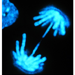
Shown below are the chromosomes within a dividing epithelial cell from a rat kangaroo. These chromosomes have been pulled to the ends of the cell which is at the end of anaphase.

The image above was used with the permission of the copyright owner, Molecular Probes..
Mitosis: Telophase

The nuclear envelope re-forms during telophase, completing the process. An overall view of the process is depicted below showing the cyclic nature of the process. Interphase is the time the cell spends between cell divisions, just doing its job in the body. Most of the time cells are in interphase.

An animated illustration of the process is shown below.
Summary of the Cell Cycle
Introduction
- Over time, many of the cells that make up our bodies age, die and need to be replaced.
- The process by which a cell reproduces to create two identical copies is known as mitosis.
- Cells formed by mitosis are known as daughter cells.
- The cell division process occurs as an orderly progression through four different stages, known collectively as the 'cell cycle'.
- Many of the abnormal traits of cancer cells are due to defects in genes that control cell division.
The Cell Cycle
- The cell cycle consists of four stages: G1, S, G2, and M.
- G1 and G2 are 'gap' phases in which the cell grows and prepares to divide.
- S in the synthesis phase in which the chromosomes (DNA) are copied (replicated).
- M is the mitotic phase in which the cell physically divides into two daughter cells.
- Most cells are NOT actively dividing. These cells are in a resting state (G).
Mitosis (M phase)
- Mitosis in normal cells produces two cells with identical genetic content.
- Mitosis has four sub-phases:
- Prophase - Chromosomes condense, the nuclear membrane breaks down, and spindle fibers form
- Metaphase - The replicated chromosomes line up in the middle of the cell
- Anaphase - Chromosomes separate and the cell becomes elongated, with distinct ends (poles)
- Telophase - Nuclear envelopes re-form at the two poles and new cell membranes are formed to create two independent cells
DNA Synthesis (S phase)
- Humans have 46 chromosomes, 23 from each parent.
- Each chromosome is comprised of a single piece of DNA containing millions of nucleotides.
- A pair of homologous chromosomes has the same genes, but can have different versions of those genes.
- In many cancer cells the number of chromosomes is altered so that there are either too many or too few chromosomes in the cells. These cells are said to be aneuploid.
- Errors may occur during the DNA replication resulting in mutations and possibly the development of cancer.
- Cells have mechanisms to correct errors due to faulty DNA replication.
- Many chemotherapy agents target the S phase of the cell cycle.
- 1 Urry, L. A., Cain, M. L., Wasserman, S. A., Minorsky, P. V., & Reece, J. B. (2017). Campbell Biology (11th ed.). Pearson.
