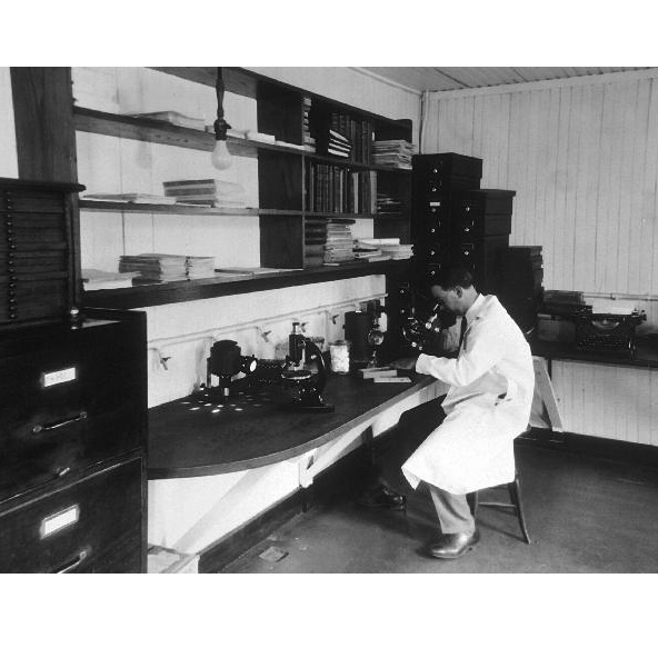
After a biopsy is taken the physician who performed the biopsy sends the specimen to a pathologist. The pathologist examines the specimens at both the macroscopic (visible with the naked eye) and microscopic (requiring magnification) levels and then send a pathology report to the physician. The report contains information about the tissue's appearance, cellular make up, and state of disease or normalcy. The pathology report is vital to the treating physician and the patient, as treatment decisions and options are made based on the information the report contains.
Sections on the following page:
- Gross or Macroscopic Report
- Microscopic Report
- Diagnoses
- FAQ Cancer Pathology
- Know the Flow: Pathology
Watch a video on breast cancer pathhology
Gross or Macroscopic Report
The first component of a pathology report is the gross or macroscopic report. This report includes the general appearance of the biopsy. Often times the pathologist will state the site from which the biopsy was taken. Also included is the shape of the tumor in question and whether or not it appears to have well-defined borders. In this section the size of the biopsy is given. Usually both the diameter or length and the weight of the specimen are given. All dimensions or size identifiers are given using the metric system of measurement. This means that lengths or diameters are given in centimeters and weights are given in grams.
NOTE: There are approximately 2.5 cm in 1 inch and 454 grams in 1 pound.
Microscopic Report
The second section of the pathology report is the microscopic report. This portion contains information and descriptions that the pathologist attains by looking under the microscope. This more technical language describes the biopsy on a cellular level. Atypical is a term used to describe cells that appear to be abnormal when examined. Several factors can define varying levels of atypia. An atypical cell often has a nucleus that is larger than usual and contains a larger amount of chromatin than is normal. Pathologists also will check the mitotic rate of the cells, which is an indication of how quickly they are multiplying. Differentiation is a term used to describe how specialized a cell is to perform a specific job in a certain tissue. The less differentiated the cell is, the more atypical it is said to be. Also, of concern in the microscopic report is whether or not it appears that all abnormal cells were removed from the biopsy site. To do so the pathologist uses the microscope to look at the borders of the biopsy. If there is a border of normal cells around the abnormal cells then the biopsy is said to have clear margins and it is assumed that all atypical cells were removed. If however there appears to be abnormal cells that lie at the edge of the removed tissue then the margins are not clear and the pathology report would contain further instruction to your physician. It would include specific information about regions that should receive further treatment, such as, additional surgery or other treatment. More on surgery and 'margins'.
Diagnosis
Normally a pathology report includes one last section, the diagnosis. In this portion the pathologist would give a technical diagnosis that would indicate whether the biopsy is benign or malignant. If it is determined that the biopsy is benign then the pathologist would most likely give insight into what level of risk the removed tissue presents to the patient's health in the future and the likeliness that this or other tumors like it would develop into more harmful malignant tumors. If it is determined that the biopsy contains malignant tissue then the pathologist would provide an indication of the cancer's severity based on findings presented in other sections of the report.
In some cases, an additional "comments" section might conclude the report that would list any other testing to be done on the biopsy and any other tests that still have incomplete results. Cancers of some organs are associated with additional specific tests. These additional tests would be included in the report.1, 2
Accordian
Pathologists use a variety of techniques to determine the stage of a tumor. Pathologists study the characteristics of cancer cells as well as the overall structure of the sample. Pathologists determine the mitotic index (how many of the cells are dividing) and the histologic 'grade', a measure of how abnormal the cells appear. One system used to stage some cancer types tumor is the TNM method, which stands for:
- Tumor - the size of the tumor
- Lymph Nodes - whether the cancer has spread to regional lymph nodes
- Metastasis - whether or not cancer has spread to other parts of the body
Learn more about cancer staging on our page dedicated to that topic.
Know the Flow is an interactive game for you to test your knowledge. To play:
- Drag the appropriate choices from the column on the right and place them in order in the boxes on the left. Note that you will only use five of the six choices to complete the game.
- When done, click on 'Check' to see how many you got correct.
- For incorrect answers, click on 'Description' to review information about the processes.
- To try again, choose 'Reset' and start over.
Please visit us on a larger screen to play this game.
- 1 College of American Pathologists [http://www.cap.org/]
- 2 The Biopsy Report: A Patient's Guide [http://www.cancerguide.org/pathology.html]
