Cancer arising in the lymphatic system, called lymphoma, is the most commonly occurring blood cancer. Approximately 1,000 people worldwide are diagnosed with lymphoma every day. The cells affected by this disease are part of the body's immune system. In 2023, the American Cancer Society estimates that 89,380 new lymphoma cases will be diagnosed and 21,080 cancer deaths due to lymphoma will occur in the United States.1
Below is a list of the information found within this section:
- Anatomy of the Lymphatic System
- Lymphoma Types
- Risk Factors
- Symptoms
- Detection and Diagnosis
- Pathology Report & Staging
- Lymphoma Tumor Biology
- Treatment
- Lymphoma Resources
- Section Summary
Anatomy Of The Lymphatic System
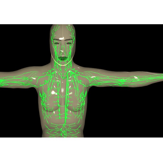
The lymphatic system is composed of a vast network of tubes (vessels) and grapelike clusters called lymph nodes. The vessels transport a colorless fluid called lymph and cells of the immune system (lymphocytes) throughout the body. The lymphatic system resembles a river system. Very thin tubes (capillaries) carry lymph into larger vessels which eventually drain into two large lymph vessels that empty into blood vessels at the base of the neck.2
The lymphatic system serves many purposes including: filtration, transport of fluid and initiation of immune responses. The vessels of the lymphatic system are responsible for absorbing and filtering the fluid which surrounds the cells and tissues of the body.2
Lymph nodes are small sac-like structures located along the lymph vessels. They are home to lymphocytes, a type of white blood cell.2 Lymph nodes store lymphocytes and help control the immune response by allowing lymphocytes to come into contact with foreign materials (antigens) in a manner that stimulates their activity.2
All lymphocytes originate from stem cells in the bone marrow but they are not all the same.2 The two main categories of lymphocytes are B cells and T cells. B cells fully develop in the bone marrow. T cells leave the bone marrow in an immature state and continue to develop in the thymus and other organs. T cells and B cells play different roles in the immune system and their functions are described more fully in the Vaccine section.2
The extensive nature of the lymphatic network allows it to serve as a way for cancer cells to spread throughout the body. Cancer metastasis frequently occurs via migration of cancer cells through the lymphatic system.2Learn more about metastasis
Types of Lymphoma
There are three types of Lymphoma:
1. Hodgkin's Lymphoma (Hodgkin's Disease)
2. Non-Hodgkin Lymphoma
3. Burkitt's Lymphoma
Hodgkin's Lymphoma
Cancer cells of these patients are usually abnormal B-cells referred to as a Reed-Sternberg (R-S) cells.3 Although less commonly observed, R-S cells may also develop from T-cells.4 R-S cells most often develop in lymph nodes located in the upper body regions and spread to neighboring lymph nodes via lymphatic vessels. There are two distinct types of Hodgkin's lymphoma: classical and non-classical Hodgkin's lymphoma.3
- Classical Hodgkin's Lymphoma: This lymphoma features R-S cells with a classical appearance. It may be diagnosed as Nodular Sclerosis Hodgkin Disease, Mixed Cellularity Hodgkin's Disease, Lymphocyte-Rich Hodgkin Disease or Lymphocyte-Depleted Hodgkin Disease.3
- Non-classical Hodgkin's Lymphoma: This lymphoma features larger cancer cells that are variants of R-S cells and is most often found in the nodes of the upper body, arms and neck.3
For more detailed information regarding the five variants of Hodgkin lymphoma, please visit the American Cancer Society web page on Hodgkin's Lymphoma.
Non-Hodgkin Lymphoma
The cancerous cells of non-Hodgkin lymphoma patients may be either T or B cells. In the United States, approximately 15% of cases of non-Hodgkin lymphoma cases develop from T lymphocytes and 85% develop from B lymphocytes.3 Normal white blood cells may develop into over thirty different variations of abnormal cells, each classified as a distinct type of non-Hodgkin lymphoma.
The American Cancer Society estimates approximately 65,540 (89%) of the 74,030 lymphoma cases diagnosed in the United States in 2010 will be classified as non-Hodgkin lymphoma.5
Some types of non-Hodgkin lymphoma include the following:
Diffuse Large B-Cell Lymphoma
Follicular Lymphoma
Mantle Cell Lymphoma
T-Cell Lymphoma
Burkitt's Lymphoma
Burkitt's lymphoma is an aggressive form of non-Hodgkin lymphoma involving B cells. It is caused by the rearrangement of parts of a chromosome including the Myc gene (translocation). Translocation disrupts Myc expression leading to abnormal cell growth and proliferation. The rate of cell division in Burkitt's lymphoma is one of the highest among human tumors. It was first described by Dr. Denis Burkitt in 1958 while he was working in Uganda. Burkitt's lymphoma was later discovered to be highly associated with the Epstein-Barr virus; this was the first time a virus was linked to a form of cancer.
The World Health Organization (WHO) describes three clinical variants of Burkitt's lymphoma: endemic, sporadic, and immunodeficiency-associated.
Endemic Burkitt's lymphoma primarily refers to cases occurring in African children. This type usually involves the facial bones, especially the jaw, maxilla, and orbit. The Epstein-Barr virus (EBV) is associated with over 90% of endemic Burkitt's lymphoma.
Sporadic Burkitt's lymphoma refers to cases occurring in no specific geographic or climatic region. This type usually involves the abdomen. Unlike endemic lymphoma, infection with EBV is found in only about 20% of sporadic lymphoma. It accounts for 40-50% of childhood non-Hodgkin lymphoma, but only 1-2% of adult lymphomas in Western Europe and the United States.
Immunodeficiency-associated Burkitt's lymphoma refers to cases occurring in patients infected with HIV, transplant patients (most often solid organ), or individuals with other immune system disorders. BL accounts for 30-40% of non-Hodgkin lymphomas diagnosed in HIV infected individuals. However, HIV is not directly related to cancer formation. EBV is found in 30-40% of these cases of Burkitt's lymphoma.6
Risk Factors
The cause of the majority of lymphoma cases is unknown. However, several factors may influence one's risk of developing lymphoma. The relative effects of these factors in any given case of cancer is variable and very difficult to determine with accuracy at this time. Some of these risk factors are discussed below.
Sex
Specific subtypes of non-Hodgkin lymphoma, such as follicular lymphoma, are predominant in women; however, non-Hodgkin lymphoma is overall more common in men. Mantle cell lymphoma shows the highest predisposition in males (70% of cases are men).7
Geography
Non-Hodgkin lymphoma is most common in developed regions of the world, specifically the United Sates, Australia, New Zealand and Europe.7 Epstein Barr virus (EBV), a type of herpes virus that infects B lymphocytes, increases a person's risk of developing fast growing lymphomas. In Africa and Southeast Asia, EBV is related to the development of Burkitt lymphoma and Hodgkin's lymphoma.3
Genetics
As discussed in the Mutation section DNA mutations can cause cancer by enhancing cell division and/or reducing tumor suppressor mechanisms. Lymphoma is rarely caused by inherited mutations in the DNA sequence and there is no increased risk of lymphoma in children of lymphoma patients.3
Age
The incidence of lymphoma peaks in individuals over 70 years of age.8 Non-Hodgkin lymphoma is rarely observed in children and most commonly develops in older adults. Less than 1% of non-Hodgkin lymphoma diagnoses reported in 2001 occurred in children under the age of 15 years.8 The age groups most frequently affected by Hodgkin's lymphoma are early adults (age 15-40) and late adults (above 55).3
Learn more about the relationship between cancer and age
Medical History
One's medical history may influence their susceptibility to the development of lymphoma.9 Individuals with autoimmune diseases are at an increased risk of developing lymphoma. Examples of diseases associated with risk of lymphoma development include: Diabetes type 1 and Rheumatoid Arthritis. 10Immunosuppressive therapies used to encourage acceptance of transplanted organs may also increase risk of lymphoma.8
Infection with certain viruses and bacteria is associated with increased risk for lymphoma. These include:
- Human immunodeficiency virus (HIV): this virus is the causative agent of AIDS.
- Epstein-Barr virus (EBV): infection with EBV is associated with an increased risk of lymphoma. In certain geographic regions, including Africa, infection with EBV is associated with Burkitt's lymphoma.
- Hepatitis C virus (HCV): The role of HCV in lymphoma risk is unclear.
- Human T-cell leukemia virus type 1(HTLV-1): This virus is associated with both lymphomas and leukemias.
- Helicobacter pylori (H. pylori): This bacteria infects the stomach and is thought to cause ulcers. Infection is also associated with an increased risk of lymphomas of the stomach.8
Symptoms
Symptoms
Lymphoma causes lymph nodes to swell. Cancerous lymph nodes are unusually swollen and may be detectable at the surface of the body. However, swollen lymph nodes may result from other causes, including infections, and do not serve as a reliable indication of cancer. Other generalized symptoms caused by lymphoma include:3
- unexplained weight loss;
- fever;
- extreme (drenching) night sweats; and
- severe "itchiness."
Specific types of lymphoma manifest themselves through different symptoms. Persons with lymphoma of the stomach or abdominal nodes may experience painful cramping, nausea, loss of appetite and constipation due to blockage of the large intestine by swollen lymph nodes. Lymphoma of the skin may be easily seen and felt. The lesions frequently appear as reddish-purple nodules directly under the skin and are often itchy.3
Detection And Diagnosis
Detection
Cancers affecting lymphocytes may be detected by several different methods. Some of these methods are discussed below:
- Blood tests: Lymphocytes spend some time in the bloodstream. If there are many lymphocytes present in the blood, this can be detected by a routine blood test, called a complete blood count or CBC. Learn more about CBC tests.
- Magnetic Resonance Imaging (MRI) has a high ability to clearly define details in the tissues surrounding the organs of the body. This method is often used to detect abnormalities and tumors in the bone marrow.11
- Skeletal Scintigraphy, or Bone Scan, is used on patients with lymphoma to assess bone damage caused by tumors. 12 In this procedure, a radioactive chemical is injected into the patient and uptake of the chemical is monitored. The use of agents that are selectively absorbed by bone produces images of the skeletal system which can be used to locate cancer.
- Computed Tomography (CT) may be used to detect lymphoma either alone or in combination with other techniques like Positron Emission Tomography.13, 11
- Positron Emission Tomography (PET) may be used to scan a person's entire body to find tumors. PET scans are helpful during treatment and help a physician to determine whether lymph tumors are malignant or benign.13, 12
Learn more about cancer diagnosis and detection.
Pathology Report And Staging
The Pathology Report
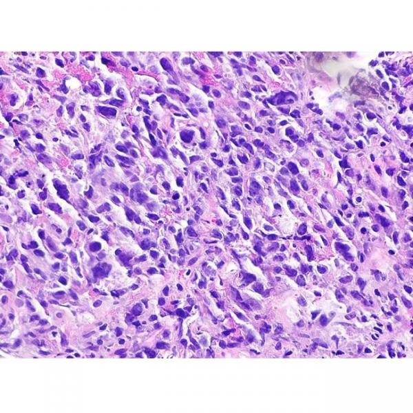
If there is suspicion that a patient may have lymphoma, a sample of tissue (biopsy) may be taken for examination. After a biopsy is taken, the physician sends the specimen to a pathologist. The pathologist examines the specimen at both the macroscopic (visible with the naked eye) and microscopic (requiring magnification) levels and then sends a pathology report to the physician. The report contains information about the tissue's appearance, cellular makeup, and state of disease or normalcy.
Learn more about the pathology report
Staging
Staging is important for identifying appropriate treatment options for particular cancer and individual. It is important to note that although the stage of a cancer is important, the prognosis may be affected by other factors, such as age of the patient and other health-related factors.
The Ann Arbor staging system is commonly used to categorize non-Hodgkin lymphoma following diagnosis. This system categorizes cancers into one of four stages. Methods to determine stage may include: biopsy, X-ray, MRI, PET and Bone Scans.3
Stages of non-Hodgkin lymphoma. The following is directly quoted or paraphrased from the sources at the end of the list (ACS and NCCN):
- Stage I: Cancerous cells are found in one lymph node or one area of a single organ outside of the lymphatic system.
- Stage II: Cancerous cells are detected in two lymph nodes or the lymph nodes of two regions of the body. Or, cancer may extend from a single group of lymph nodes to surrounding organs.
- Stage III: Lymph nodes on both sides of the diaphragm contain tumor cells.
- Stage IV: Lymphoma has spread out of the lymphatic system into organs that are not situated adjacent to a diseased node.3, 14
Learn more about cancer staging
Tumor Biology
Genetic changes that occur in cancer include mutation of key regulatory genes, changes in protein products, and changes in the amount of product produced by genes (gene expression). As changes accumulate, cells become more abnormal and cancer progresses. Details about the genetic changes associated with cancer can be found in the Mutation section. Some of the genetic elements shown to be important in the development of lymphoma are discussed below.
bcl-6
The bcl-6 protein is a transcription factor, It binds to DNA and alters the activity of specific genes. The bcl-6 protein helps regulate the growth and development of B cells.15
Alterations in this gene, due to base mutations and chromosomal abnormalities, is thought to play a role in the development of many cases of lymphoma.15, 16
p53
p53 is a tumor suppressor gene whose protein is responsible for the regulation of cell division (and cell death) in normal cells. For at least some patients, mutation of the p53 gene is thought to contribute to the unregulated cell growth seen in Hodgkin's lymphoma.17
Learn more about abnormal p53 and cancer development
HDM2
HDM2 is a protein that regulates the activity of the p53 protein. In normal cells, HDM2 only interferes with p53 when needed. In lymphoma development, however, mutations of the HDM2 gene may cause continuous production of the HDM2 protein. The protein continuously prevents p53 from suppressing cell division, contributing to the uncontrolled proliferation of the cancer cells.18
Treatment
Specific treatment plans for lymphoma depend on the type and stage of the disease. As our focus is on the biology of the cancers and their treatments, we do not give detailed treatment guidelines. Instead, we link to organizations in the U.S. that generate the treatment guidelines.
The National Comprehensive Cancer Network (NCCN) lists the following treatments for non-Hodgkin's lymphoma:
- Surgery: For more details about this type of treatment view the section on Surgery.
- Radiation Therapy: Radiation is used to treat disease confined to one area of the body, often seen in patients with stage I or II lymphoma. Radiotherapy uses high energy rays to kill lymphoma cells. For more details view the section on Radiation Therapy
- Chemotherapy: Often several chemotherapy drugs are given in combination. A combination commonly administered used to treat non-Hodgkin lymphoma is known as C.H.O.P., which includes cyclophosphamide (Cytoxan®, or Neosar®), doxorubicin or hydoxydoxorubicin (Adriamycin®), vincristine (Oncovin®) and prednisone.3For more details on this treatment view the section on Chemotherapy.
- Antibody-based treatments: Antibodies are injected into the patient and recognize, bind and destroy lymphoma cells.3 Rituximab (Rituxan®) is a human-mouse hybrid antibody that may be administered to lymphoma patients. It binds to a specific subset of B lymphocytes and causes their death.4 Rituximab may be used alone or in conjunction with chemotherapy. For more details on this method view the section on Antibody-based treatment. Learn more about this drug from the manufacturer here.
- Stem Cell Transplantation: For details on this treatment view the section on Stem Cell Transplantation.
The NCCN also lists chemotherapy and radiation as treatments for Hodgkin's lymphoma.
The National Cancer Institute website contains information about treatments for Childhood Hodgkin's Lymphoma and Adult Hodgkin's Lymphoma.
For more information about how these and other cancer treatments work, refer to the Cancer Treatments section.
Information about clinical trials:
- General clinical trial information from CancerQuest
- Click here for information about clinical trials from the National Cancer Institute.
- Click here for information about clinical trials from Georgia Clinical Trials Online.
Lymphoma Resources
Risks for Lymphoma
Hodgkins Lymphoma Risk Factors
Non-Hodgkin Lymphoma Risk Factors
Detection and Diagnosis of Lymphoma
Winship Cancer Insitute: Lung Cancer Diagnosis and Staging Make an Appointment
Winship Cancer Institute: Lymphoma Cancer Diagnosis and Staging Make an Appointment
What You Need to Know About: Lymphoma (NCI)
Lymphoma Treatments
Adult Hodgkin Lymphoma Treatment
Adult Non-Hodgkin Lymphoma Treatment
AIDS-Related Lymphoma Treatment
Childhood Hodgkin Lymphoma Treatment
Childhood Non-Hodgkin Lymphoma Treatment
Lymphoma Survivorship
Long Term Risks for Lymphoma Survivors
International Lymphoma Resources
The Leukemia and Lymphoma Society of Canada
Macmillan Cancer Support: Lymphoma (UK)
We Care Health Services: Lymphoma (India)
Cancer Council Australia: Lymphoma
Section Summary
Introduction
- Lymphoma is the most commonly occurring blood cancer in the U.S.
- Lymphoma is a cancer of white blood cells that arises in the lymphatic system.
Types of Lymphoma
- Hodgkin's lymphoma frequently develops from B-cells, but may also develop from T-cells.
- The majority of lymphoma cases are non-Hodgkin lymphoma
Risk Factors
- Non-Hodgkin lymphoma is more common in developed regions of the world and in men.
- Infection with the Epstein Barr virus and other such viruses increases the risk of developing lymphoma.
Symptoms
- Swollen lymph nodes, unexplained weight loss, fever, extreme night sweats, and severe "itchiness".
- Symptoms vary with the location in which the cancer arises.
Detection and Diagnosis
- Lymphatic tumors may be detected by an MRI, a bone scan, a CT scan, or a PET scan.
Pathology Report and Staging
- A pathologist examines a tissue sample micro- and macroscopically in order to create a pathology report.
- The Ann Arbor staging system is commonly used to categorize non-Hodgkin lymphoma into one of four stages.
Lymphoma Tumor Biology
- Many genetic changes occur in cancer. Details can be found in the Mutation section.
- BCL-6, p53, and HDM2 are all genes involved in the development of lymphoma.
Treatment
- Specific treatment plans for lymphoma depend on the type and stage of disease.
- Non-Hodgkin's lymphoma can be treated with surgery, radiation therapy, chemotherapy, stem cell transplantation, or targeted treatments.
- Hodgkin's disease is treated with chemotherapy, radiation and bone marrow transplantation.
- 1 American Cancer Society.Cancer Facts & Figures 2023. Atlanta: American Cancer Society. (2023). Retrieved from https://www.cancer.org/research/cancer-facts-statistics/all-cancer-facts-figures/2023-cancer-facts-figures.html
- 2abcdefg Vander, Arthur J., James H. Sherman, Dorothy S. Luciano. Human Physiology, 6th Edition. McGraw-Hill, Inc. NY, NY (1994).
- 3abcdefghijklmn Learn About Lymphoma, Non-Hodgkin's type. American Cancer Society. Accessed October 2010. [http://www.cancer.org/cancer/non-hodgkinlymphoma/index]
- 4ab Long J, Versea L. "Treatment approaches and nursing considerations for non-Hodgkin's lymphoma." Semin Oncol Nurs. (2006 May);22(2):97-106. [PUBMED]
- 5 Cancer Facts and Figures 2010. American Cancer Society. [http://www.cancerquest.org/sites/default/files/assets/pdf/facts%26figures2010.pdf]
- 6 Jonathan L. Hecht, Jon C. Aster. Molecular Biology of Burkitts Lymphoma. Journal Clinical Oncology. 2000; 18(21):3707-3721. [PUBMED]
- 7ab Ekstrom-Smedby K. "Epidemiology and etiology of non-Hodgkin lymphoma--a review." Acta Oncol. (2006);45(3):258-71 [PUBMED]
- 8abcd Grulich AE, Vajdic CM. "The epidemiology of non-Hodgkin lymphoma." Pathology (2005 Dec);37(6):409-19. [PUBMED]
- 9 Landgren O, Bjorkholm M, Montgomery SM, Hjalgrim H, Sjoberg J, Goldin LR, Askling J. "Personal and family history of autoimmune diabetes mellitus and susceptibility to young-adult-onset Hodgkin lymphoma." Int J Cancer (2006 Jan 15);118(2):229-52 [PUBMED]
- 10 Landgren O, Engels EA, Pfeiffer RM, Gridley G, Mellemkjaer L, Olsen JH, Kerstann KF, Wheeler W, Hemminki K, Linet MS, Goldin LR. "Autoimmunity and susceptibility to Hodgkin lymphoma: a population-based case-control study in Scandinavia." J Natl Cancer Inst (2006 Sep 20);98(18):1321-30. [PUBMED]
- 11ab Schmidt GP, Schoenberg SO, Reiser MF, Baur-Melnyk A. "Whole-body MR imaging of bone marrow." Eur J Radiol (2005 Jul);55(1):33-40. [PUBMED]
- 12ab Sheth S, Ali S, Fishman E. "Imaging of renal lymphoma: patterns of disease with pathologic correlation." Radiographics (2006 Jul-Aug);26(4):1151-68. [PUBMED]
- 13ab Hicks RJ, MacManus MP, Seymour JF. Initial staging of lymphoma with positron emission tomography and computer tomography. (2005) Seminars in Nuclear Medicine. 35(3):165-75. [PUBMED]
- 14 National Comprehensive Cancer Network Website: Non-Hogkin's Lymphoma Stages. Accessed 2 June 2010. [http://www.nccn.org/patients/patient_gls/_english/_non_hodgkins/4_stages.asp]
- 15ab Migliazza A, Martinotti S, Chen W, Fusco C, Ye BH, Knowles DM, Offit K, Chaganti RS, Dalla-Favera R. "Frequent somatic hypermutation of the 5' noncoding region of the BCL6 gene in B-cell lymphoma." Proc Natl Acad Sci USA (1995 Dec 19);92(26):12520-4. [PUBMED]
- 16 Keller CE, Nandula S, Vakiani E, Alobeid B, Murty VV, Bhagat G. "Intrachromosomal rearrangement of chromosome 3q27: an under recognized mechanism of BCL6 translocation in B-cell non-Hodgkin lymphoma." Hum Path (2006 Aug);37(8):1093-9. [PUBMED]
- 17 Feuerborn A, Moritz C, Von Bonin F, Dobbelstein M, Trumper L, Sturzenhofecker B, Kube D. "Dysfunctional p53 deletion mutants in cell lines derived from Hodgkin's lymphoma." Leuk Lymphoma (2006 Sep);47(9):1932-40 [PUBMED]
- 18 Sanchez-Aguilera A, Garcia JF, Sanchez-Beato M, Piris MA. "Hodgkin's lymphoma cells express alternatively spliced forms of HDM2 with multiple effects on cell cycle control." Oncogene (2006 Apr 27);25(18):2565-74 [PUBMED]